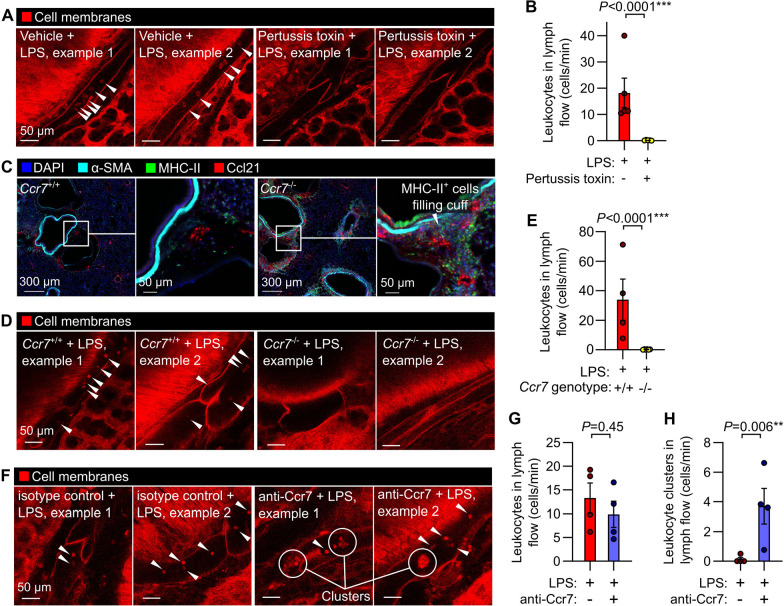
Figure 4: Interventions targeting chemokine receptor signaling alter leukocyte trafficking through lung lymphatics.
(A) Leukocyte flow through pulmonary lymphatics from Rosa26mTmG mice treated with either vehicle control or pertussis toxin before challenge with LPS with (B) quantification of effect of pertussis toxin. (C) Immunofluorescence images of Ccr7+/+ wild-type mice and Ccr7−/− knockouts showing accumulation of MHC-II+ cells in bronchovascular cuffs. (D) Pulmonary lymphatic leukocyte flow in LPS-treated Ccr7+/+:Rosa26mTmG mice and absence of intralymphatic leukocytes in Ccr7−/−:Rosa26mTmG mice, with (E) quantification of effect of Ccr7 knockout. (F) Pulmonary lymphatics in LPS-treated Rosa26mTmG mice given either isotype-matched control antibody or anti-Ccr7 by oropharyngeal aspiration together with LPS, with (G) absence of effect of antibody on total leukocyte flow and (H) quantification of flow of clusters of leukocytes in lung lymph. White arrowheads indicate intralymphatic leukocytes, circles highlight intralymphatic leukocyte clusters. Graphs show means ± SEM. P-values are from unpaired, two tailed t-tests on log10-transformed datasets. Group sizes: (B) n=5; (E, G, H) n=4.

