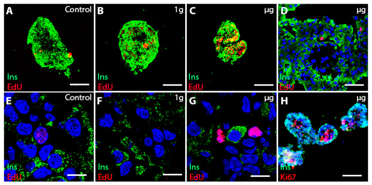
Figure 3.
Free-floating islets alone in ground control, 1 g, and µg conditions. Upper panel: insulin + cells (green insulin, red EdU, or Ki67 (H) in the non-fused islets (A–C) proliferate in µg conditions) (C). The majority of islets are fused (D,H). Lower panel: confocal images showing EdU labeling in µg also in insulin-control (E), 1 g (F) and µg (G) groups and insulin + cells (H) in µg group. Bar (A–C) = 100 µm; bar (D) = 30 µm; bar (E–G) = 10 µm; bar (H) = 150 µm. Nuclei were stained blue with Hoechst.

