Figure 7.
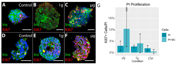
A: Overview of 3D-printed islets alone (upper panel) and PI+BC (lower panel) in control (A,D), 1 g (B,E), and µg (C,F). The proliferation of islet cells was detected three weeks after the space voyage, only in µg-exposed islets (C,F). Proliferating cells (red Ki67) are insulin-positive (green) in islet-alone cultures (upper panel, right) and insulin-negative in co-culture (lower panel, right). (G): Graph shows increased proliferation of islet cells after µg exposure in 3D-printed scaffolds in islets alone and PI+BC co-cultures compared to control groups. Section signs denote statistically significant (§—p < 0.05) differences between the specified and equivalent ground control groups. Bar = 100 µm. Nuclei stained blue with Hoechst.

