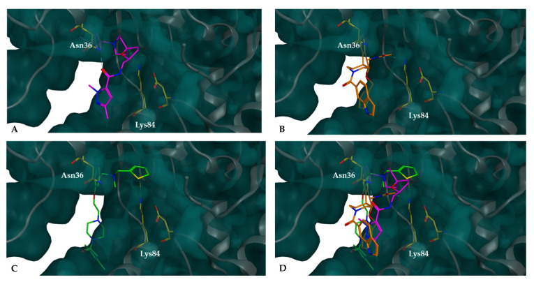
Figure 7.
Compounds docked into the binding pocket of the XPG protein are represented as follows: XPG highlighting the Lys84 and Asn36 residues inside the binding pocket; (A) docked complexes of XPG with potential inhibitor CB41-G (purple), (B) docked complexes of XPG with potential inhibitor CB60-G (orange), (C) docked complexes of XPG with potential inhibitor CB22-G (green), and (D) overlap of the three small molecules. All these molecules fit snugly within the pocket, effectively blocking access to the top of the cavity.

