Figure 5.
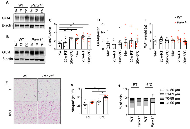
Cold-induced WAT morphological changes are amplified in Panx1−/− mice. (A,B) Illustrative Western blots (Western blot original images can be found in Supplementary Materials) and (C,D) quantification of Glut4 expression in WAT (C) and skeletal muscle (D) from 14-week-old or 20-week-old WT and Panx1−/− mice housed either at 22 °C or at 6 °C during 4 weeks. Glut4 expression was normalized towards beta actin. (E) Weight of WAT pads from 14-week-old or 20-week-old WT and Panx1−/− mice at 22 °C or exposed to cold. (F) Hematoxylin and eosin staining on paraffin sections from WAT of WT and Panx1−/− mice at 22 °C or exposed to cold. Quantification of the numbers (G) of adipocytes in the WAT of WT and Panx1−/− mice at 22 °C or exposed to cold. Data are shown as individual data points and expressed as mean ± SEM. (H) Quantification of the proportion of small (≤50 μm), medium (51–69 μm), large (70–89 μm), and very large (≥90 μm) adipocytes in WAT of WT and Panx1−/− mice at 22 °C or exposed to cold. * p ≤ 0.05 and ‡ p ≤ 0.001.

