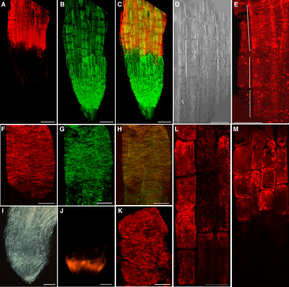
Figure 7.
COB Immunolocalization in 5-d-Old Roots.
Whole-mount confocal scanning micrographs of wild-type roots ([A] to [E]), wild-type elongating cells ([F] to [H] and [L]), and ton2 roots ([I] to [K]). Bars = 50 μm in (A) to (E) and (I), (J), (L) and (M) and 10 μm in (F) to (H) and (K). Brackets in (D) and (E) indicate the same three elongating cells.
(A), (E), (F), and (J) to (L) Indirect fluorescence immunolocalization of COB using anti-COB antibodies.
(B) and (G) Visualization of cortical microtubules by indirect immunofluorescence.
(C) Combined images of COB (A) and microtubules (B) at the root tip.
(D) and (I) Differential interference contrast micrographs of the wild-type elongation zone and the ton2-14 root tip, respectively.
(H) Combined images of COB (F) and microtubules (G) in an elongating cell.
(L) COB staining after treatment with 10 μM oryzalin for 45 min. Note patches, compared with the normal banding pattern.
(M) COB staining after treatment with 50 μM brefeldin A for 45 min. Note intracellular accumulation of COB-containing clumps.

