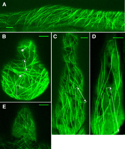
Figure 1.
Visualization of Cortical Microtubules in L. japonicus Plants Transformed with GFP-TUA6.
(A) Root epidermal cell.
(B) Emerging (Zone I) root hair cell. Asterisks indicate the sites of microtubule nucleation.
(C) Growing (Zone II) root hair cell.
(D) Mature (Zone III) root hair cell.
(E) Emerging (Zone I) root hair cell of 10-d-old Lotus plant.
Sample measurements of microtubule length ([B] to [D], double-sided arrows) and angles ([B], arrow). Bars = 5 μm.

