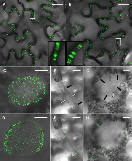
Figure 7.
Stable Expression of AtRGP2:GFP and of MPTMV:GFP in Transgenic Tobacco Plants.
Identical fluorescence patterns are presented by leaf epidermal cells in AtRGP2:GFP (A) and MPTMV:GFP (B) expressing transgenic plants: punctate fluorescence spans walls as elongated bars, or paired foci, or appears as single foci inside the cell wall. The areas inside the white boxes are enlarged in the insets in (A) and (B). The trichome-epidermis interface in AtRGP2:GFP (D) and MPTMV:GFP (C) expressing plants displays identical fluorescence patterns. In trichome (F) and spongy mesophyll cells (H) of AtRGP2:GFP expressing transgenic tobacco, fluorescence is detected only in wall areas where there is cell–cell contact and is absent from wall areas without cell–cell contact. To emphasize wall partitions, the same trichome (E) and spongy mesophyll cells (G) are shown with the fluorescence channel turned off. Black arrows indicate wall areas where there is cell–cell contact, and white arrows indicate wall areas without cell–cell contact. Bars = 20 μm.

