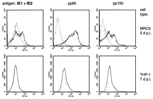
FIG. 4.
Expression of lytic-phase antigens in HCMV-infected THP-1 cells at day 7 p.i. The top panels show immunocytometric histograms of HCMV-infected MRC5 fibroblasts (total cell count = 10,000) at day 2 p.i. The lower panels show histograms of infected THP-1 cells (total cell count = 500,000) at day 7 p.i. FL1-H, relative intensity of fluorescence. Dotted lines represent uninfected cells.

