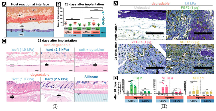
Figure 5.
(I) PEG-GAG hydrogels implanted subdermally. (A) Scheme of the implantation site: Dermis (De), Adipose Tissue (AdTi), Skeletal muscle (SkMu), Hypodermis (HyDe), Capsule (Cap), Implant (Imp). (B) Foreign body reaction thickness, ns—not significant, ***—p ≤ 0.001. (C) Immunostaining after 28 days. Scale bar 200 μm. (II) Angiogenic effects, hydrogels loaded with different cytokines. (A) CD31 immunostaining of hydrogels (dark blue) with vascular structures (brown). Scale bar 200 μm. (B) Vessel formation [125], ns—not significant, ***—p ≤ 0.001.

