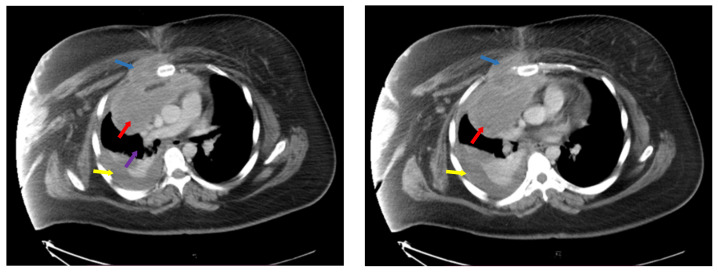
Figure 1.
Computer tomography at diagnosis showing mediastinal adenopathy block (red arrows), with compression of vasculature (purple arrow) and invasion of thoracic wall and right pectoral muscle (blue arrows), pleural effusion (yellow arrows). Left side—ce reprezinta (descriere), Right side—ce reprezinta (descriere).

