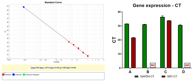
Figure 3.
SRY gene DNA quantification in gDNA via RT PCR. The red dots are the standard diluted samples, starting from 50 ng gDNA. The blue dots are the two positive SRY samples from patient in duplicate samples. A—Male control sample (50 ng/reaction); B—Female control sample (50 ng/reaction), C—Patient at 34 weeks of pregnancy (50 ng/reaction), D—Patient after 4 years (50 ng/reaction), ND—not detected. Left side—image depicting calibration curve and linearity for SRY determination, Right side—graphic depicting the comparison between gene amplification for SRY and housekeeping gene GAPDH (with the maximum determination at cycle 40).

