FIG. 2.
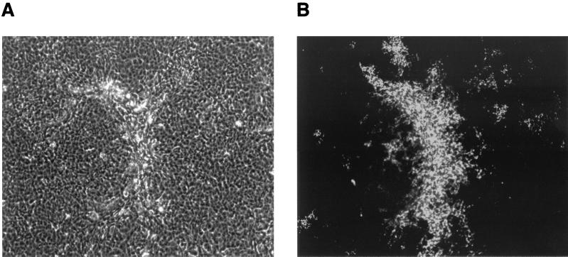
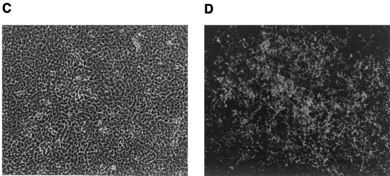
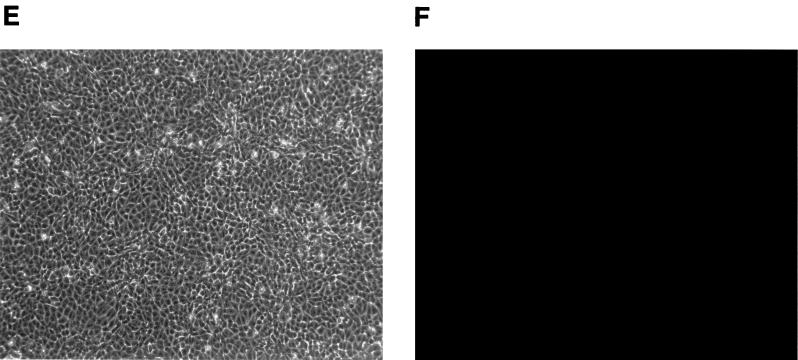
MCF13 MLV infection of mink epithelial cells produces cytopathic foci. Ten thousand CCL64 mink epithelial cells were infected with MCF13 MLV (A and B) or 4070A amphotropic MLV (C and D) at an MOI of 1. Cells were also mock infected with medium (E and F). Six days after virus infection, cells were examined by phase microscopy (A, C, and E) and fluorescence microscopy (B, D, and F). Fluorescence assays were performed by first staining cells with MAb 83A25, a monoclonal antibody that recognizes nearly all MLV glycoproteins, and subsequently with an FITC-labeled secondary anti-rat antibody.


