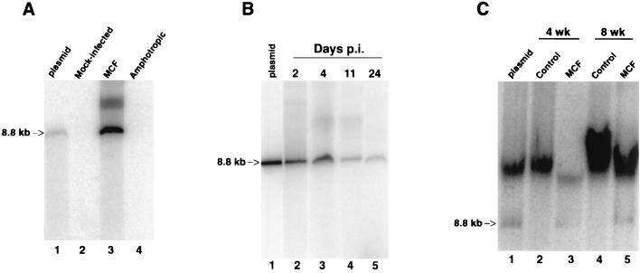
FIG. 5.
MCF13 MLV infection produces high levels of unintegrated linear viral DNA. Southern blot analysis was performed on Hirt-extracted DNA from mink epithelial cells (A and B) and thymic lymphocytes from virus-inoculated mice (C). (A) Hirt-extracted DNA from mink cells that were either mock infected (lane 2) or infected with MCF13 MLV (lane 3) or with 4070A amphotropic MLV (lane 4) after 6 days p.i. Plasmid DNA (2.5 ng) corresponding to the genomic-length MCF13 MLV was electrophoresed in lane 1. DNA was hybridized with a 32P-labeled 1.6-kb DNA fragment corresponding to MCF13 MLV gag sequences. (B) Mink cells were infected with MCF13 MLV at an MOI of 1 for 2 days (lane 2), 4 days (lane 3), 11 days (lane 4), and 24 days (lane 5). Lane 1 contains 10 ng of linear plasmid MCF13 MLV genomic-length DNA. A 32P-labeled probe consisting of 8.2 kb of MCF13 MLV genomic DNA was used for hybridization. (C) Hirt DNA was prepared from thymic lymphocytes which were isolated from thymuses of either uninoculated age-matched control AKR mice (lanes 2 and 4) or MCF13 MLV-inoculated mice (lanes 3 and 5) at 4 and 8 weeks postinoculation of virus. Lane 1 contains 2.5 ng of linear plasmid MCF13 MLV DNA and Hirt DNA from a control mouse. DNA was hybridized with a 32P-labeled probe consisting of 8.2-kb MCF13 MLV DNA. Arrows, 8.8-kb linear MCF13 MLV DNA.

