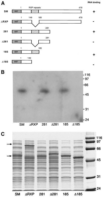
FIG. 1.
Analysis of in vitro RNA binding by SM mutants. (A) Structure of GST-SM fusion proteins. GST fused to the amino terminus of SM mutants is shown in gray. Amino acids at sites of deletion or truncation are shown above each diagram. The arginine-rich region (RXP repeats) is shown with diagonal lines. Potential cleavage sites in the arginine-rich region are depicted by the wavy line. (B) Northwestern assay. Lysates of bacteria expressing full-length or mutant SM fusion proteins were electrophoresed, transferred to polyvinylidene difluoride membranes, and probed with single-stranded radioactively labeled CAT RNA probe. Molecular masses are shown at right in kilodaltons. (C) Coomassie-stained gel of proteins analyzed by Northwestern assay in panel B. Locations of full-length GST-SM (upper arrow) and its major cleavage product (lower arrow) are shown at left. Molecular masses are shown at right in kilodaltons.

