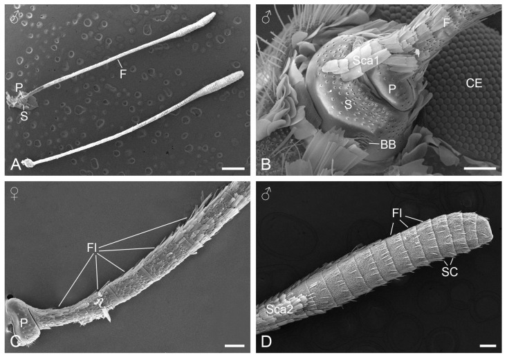
Figure 1.
SEM micrographs of Pseudozizeeria maha antennae. (A) A pair of antennae. (B) Basal region of the antenna. (C) Pedicel and proximal flagellomeres. (D) Distal flagellomeres forming the antennal club. BB, Böhm’s bristle; CE, compound eye; F, flagellum; Fl, flagellomere; P, pedicel; S, scape; SC, sensillium chaeticum; Sca1, scale with pores; Sca2, scale without pores. Scale bars: (A) = 600 µm; (B,C) = 60 µm; (D) = 100 µm.

