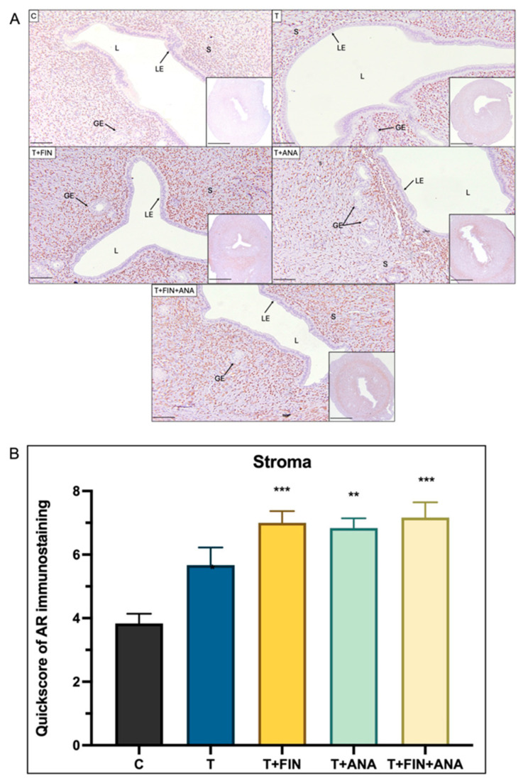
Figure 2.
Distribution of the androgen receptor (AR) in the uterus of rats. (A) The dark brown colouring, indicated by arrows, indicates the antibody binding site of the AR, which appears to be located in the stroma. Strong dark brown staining was observed in all groups only in the stroma, but not in the epithelial gland and the glandular epithelium. (B) Semi-quantitative evaluation of AR immunostaining for the stroma. C: normal control; T: testosterone propionate; T + FIN: testosterone propionate + finasteride; T + ANA: testosterone propionate + anastrozole; T + FIN + ANA: testosterone propionate + finasteride + anastrozole. LE: luminal epithelium; GE: glandular epithelium; L: endometrial lumen; S: stroma. Scale bar = 200 μm, scale bar inset = 1 mm, magnification 40× and 200×. Error bars represent standard error of the mean (SEM). n = 6 in each group, ** p < 0.01, *** p < 0.001 significance compared to normal control group.

