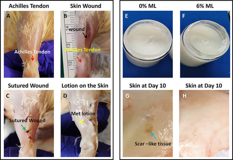
Fig 1. Photographs show the appearance of ML and rat skin over the Achilles tendon before and after surgery.
A: The rat skin over the Achilles tendon (red arrow labeled area) was prepared for surgery after removing the hair by a shaver. B: A wound (2 cm) was created in the skin over the Achilles tendon (black arrow labeled area). C: The skin wound was sutured (green arrow labeled area). D: The ML was applied on the sutured skin wound area (yellow arrow labeled material). E: The appearance of the 0% ML shows a white cream-like product. F: The appearance of the 6% ML shows a white cream-like product. G: Gross inspection shows some scar-like tissue appeared in the inside of the skin over the Achilles tendon in 0%ML. H: Gross inspection shows some scar-like tissue appeared in the inside of the skin over the Achilles tendon in 6%ML.

