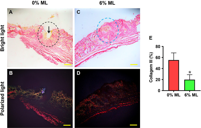
Fig 6. Metformin lotion decreases collagen III levels in the wounded skin of rats after 10 days of wound healing.
Under a bright-field microscope, a large gap (black arrow in A) is found in the 0% ML treated wound areas (black dashed line in A), while no gap is found in the wound area treated with 6% ML (blue dashed line in C). Under polarized light microscopy, high levels of collagen III are found in the 0% ML treated wound area (green fluorescence in B), whereas the wound area treated with 6% ML is positively stained for collagen I (red fluorescence in D). Semi-quantification confirms these results (E). *p < 0.001, compared to the 0% ML treated skin wound. ML: Metformin Lotion. Scale bars: 500 μm (yellow). Analysis was conducted using Picro-Sirius Red staining.

