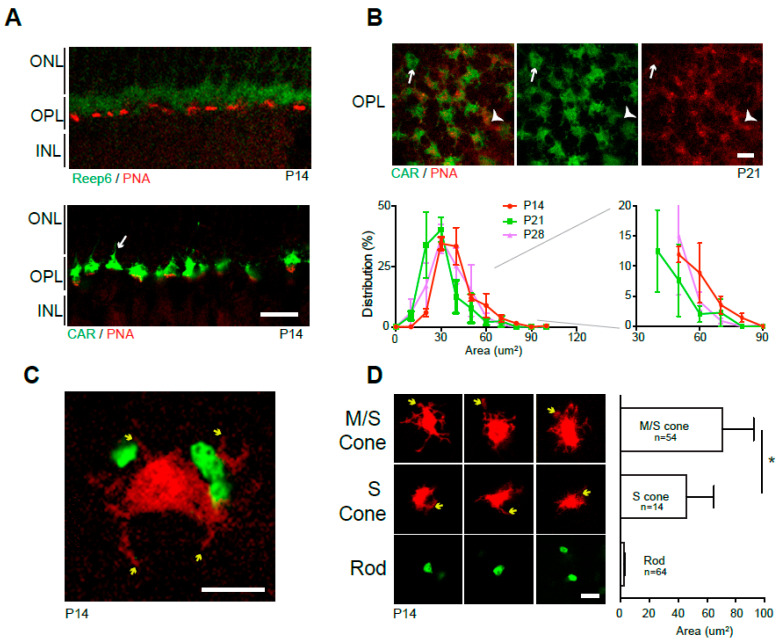
Figure 2.
Wild-type spherule and pedicle. (A) P14 vertical retina sections stained by anti-Reep6 (spherules; green) and PNA (pedicles; red) (upper panel) or anti-CAR (M-cone, green) and PNA (S-cone, red) (lower panel). (B) Horizontal OPL images of retina whole mounts stained by anti-CAR and PNA (upper panels). Pure M-cone (arrows) and S-cone (arrowheads) pedicles are observed. The graph shows the distribution (%, average ± SEM) of CAR pedicle areas in OPL of P14, 21, and 28 whole mount retinas. Over 180 CAR positive pedicles were measured from 3 wild-type C57BL/6J retinas. The area distribution after 30 μm2 is magnified on the left side. (C) Spherules (green) and a pedicle (red) in CD1 retina whole mount labeled by in vivo electroporation of Nrlp-EGFP and S-opsinp-tdT. Telodendria (yellow arrows) are observed. (D) Representative images of M/S-, S-cone pedicles and spherules and their area size comparison. Telodendria (yellow arrows) are observed in cones. M-cone and pure S-cone pedicles are segregated by anti-M-opsin staining in the retina whole mounts labeled by S-opsinp-tdT electroporation. M/S-pedicles (n = 54), S-pedicles (n = 14), and Rod spherules (n = 64) from 3 to 5 wild-type CD1 retinas were measured. The graph displays the average ± SD of each: 70.79 ± 21.48 μm2 for M pedicles, 45.91 ± 18.97 μm2 for S pedicles, and 2.64 ± 0.81 μm2 for rod spherules. * p ≤ 0.05, two-tailed T-test. Abbreviations: CAR, cone arrestin; PNA, peanut agglutin lectin; S-opsin promoter-driven tdTomato (S-opsinp-tdT); P, postnatal day; ONL, outer nuclear layer; OPL, outer plexiform layer; INL, inner nuclear layer. Scale bars, 10 μm in (A), and 5 μm in (B–D).

