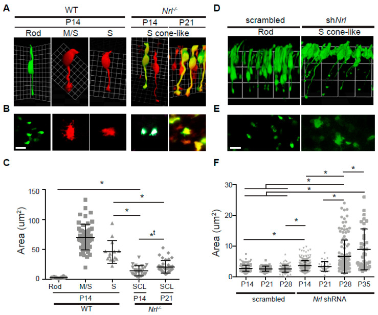
Figure 4.
Pre-synapse comparison in wild-type rod, M/S-cone, pure S-cone, and Nrl-/- or Nrl knockdown S-cone-like (SCL) photoreceptors. (A) Representative Volocity 3D images of wild-type rod, M/S-cone, pure S-cone, and Nrl-/- SCL photoreceptors, taken from wild-type or Nrl-/- retina whole mounts labeled by Nrlp-EGFP, S-opsinp-tdT. Rods (green only), cones (red only) and SCLs (mixed green and red) were imaged. M/S- and pure S-cones were differentiated by staining with an anti-M-opsin antibody. (B) Representative confocal images of pre-synapse terminals of wild-type rod, M/S-cone, pure S-cone, and Nrl-/- SCL photoreceptors. (C) Size distribution of pre-synapses in wild-type rod (n = 64), M/S-cone (n = 54), pure S-cone (n = 14) and Nrl-/- SCL (P14, n = 25; P21, n = 38) photoreceptors. (D) Representative Volocity 3D images of P28 retina whole mounts labeled by electroporation of scrambled or Nrl shRNA plasmid (shNrl) with Nrlp-EGFP (2:1 ratio). (E) Representative confocal images of pre-synaptic terminals expressing scrambled or Nrl shRNA. (F) Size distribution of pre-synapses in control (P14, n = 248; P21, n = 64; P28, n = 98) and developing Nrl shRNA SCL photoreceptors (P14, n = 246; P21, n = 31; P28, n = 124; P35, n = 36) labeled with Nrlp-EGFP. Data of area in measurement were analyzed by one-way ANOVA (Tukey or Kriskal–Wallis test) and T-test (two-tailed) in Prism. *, statistically meaningful in one-way ANOVA and T-test; *t, statistically meaningful in T-test. Abbreviations: WT, wild-type; Nrl, neural retina leucine zipper; 3D, three-dimensional; Nrlp-EGFP, Nrl promoter-driven enhanced GFP; S-opsin promoter-driven tdTomato (S-opsinp-tdT); P, postnatal day; SCL, S-cone-like; shRNA, short hairpin ribonucleic acid. Scale bars, 5 μm in (B,E).

