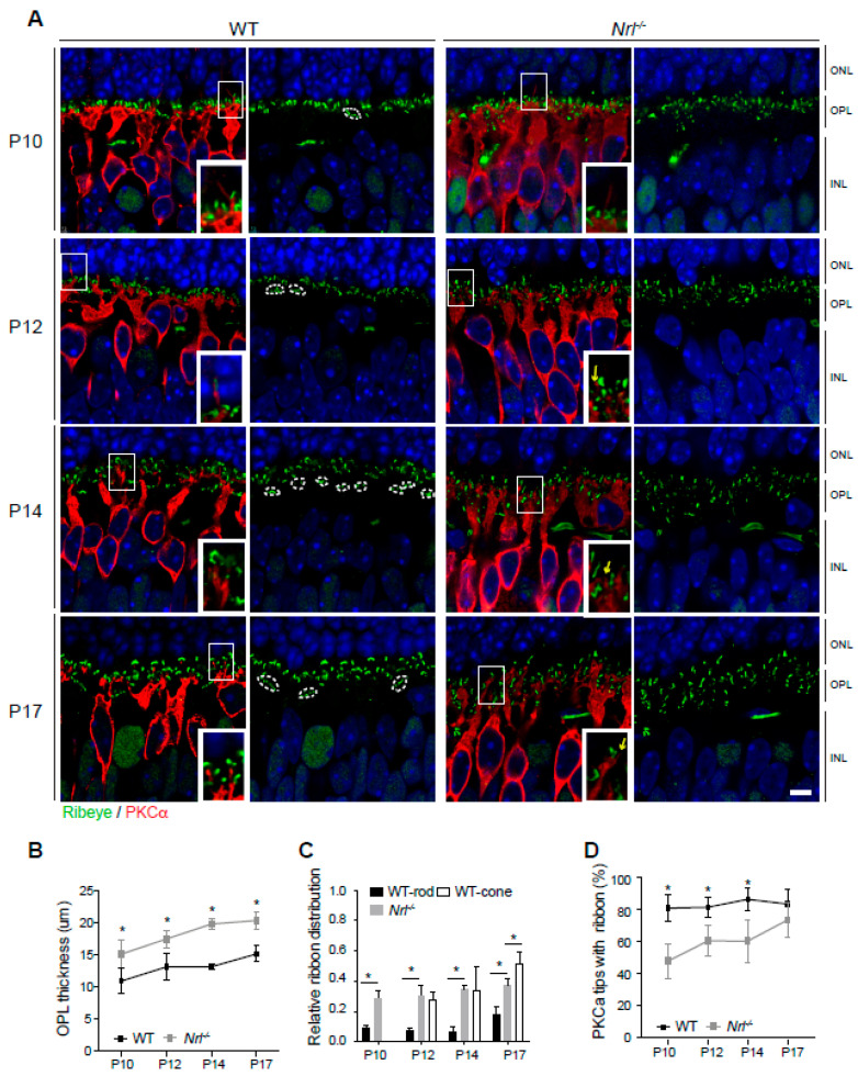
Figure 5.
Outer plexiform layer development and synaptic connection in Nrl-/- retina. (A) Developing (P10 to P17) retinas of wild-type and Nrl-/- mice stained by anti-Ribeye (green) and anti-PKCα (red). Clustered pedicle ribbons (white dotted lines) and dendritic tips of rod bipolar neurons without synaptic ribbons (yellow arrows) are observed. (B) Comparison of OPL thickness in developing wild-type and Nrl-/- retinas. Measurement was quantified on five images of the middle retina (with optic nerve head) from each of three to four animals in different developing stages. Values represent mean ± SD. * p ≤ 0.05, two-tailed T-test. (C) Comparison of the ribbon distribution in OPL. Distance of ribbon location from the ONL bottom when the OPL thickness is considered 1.0. The location of individual ribbons was measured with each OPL thickness in over two images from each of three to four animals. Values represent mean ± SD. * p ≤ 0.05, two-tailed T-test. (D) Number comparison (%) of rod bipolar neuron dendritic tips with or without ribbons aligned at their tops. Dendritic tips of rod bipolar neurons were measured at P10 (WT, n = 363; Nrl-/-, n = 627), P12 (WT, n = 445; Nrl-/-, n = 953), P14 (WT, n = 433; Nrl-/-, n = 691), and P17 (WT, n = 197; Nrl-/-, n = 269). Values represent mean ± SD. * p ≤ 0.05, two-tailed T-test. Abbreviations: WT, wild-type; Nrl, neural retina leucine zipper; P, postnatal day; PKCα, Protein Kinase C alpha; ONL, outer nuclear layer; OPL, outer plexiform layer; INL, inner nuclear layer. Scale bars, 5 μm in (A).

