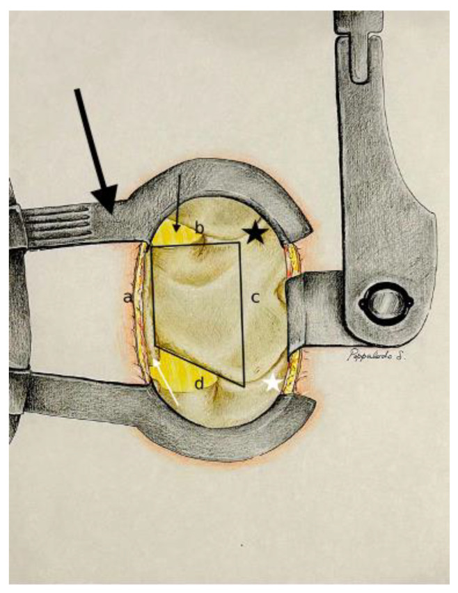
Figure 1.
Exposition after Caspar distractor position and skeletalization of homolateral spinous process and homolateral lamina (Step 1). Dentification of the posterior surgical trapezoid: (a) from caudal to cranial point of the base of the spinous process; (b) from the cranial point of the base of the spinous process to the medial third of the superior articular process (black star); (c) from the medial third of the superior articular process to the medial third of the inferior articular process (white star); (d) from the caudal point of the base of the spinous process to the medial third of the inferior articular process. Base of the spinous process (white arrow); yellow ligament (black arrow); Caspar distractor (big black arrow).

