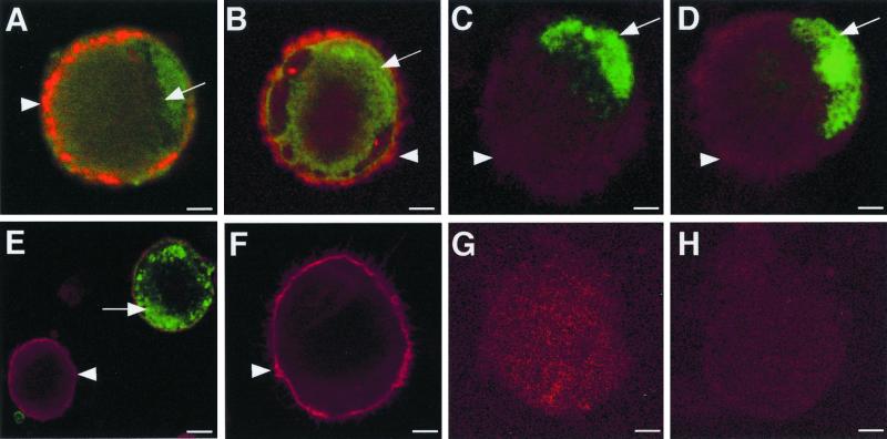
FIG. 2.
Immunofluorescence staining of CD1a and VZV antigens in VZV-infected DCs. Two days postinfection, DCs infected with VZV strain Schenke (A to E, G) and mock infected (F and H) were incubated with a mouse monoclonal antibody to CD1a (A to F) and rabbit polyclonal antibodies to ORF62 (A and F), ORF4 (B), ORF29 (C), ORF61 (D), and glycoprotein C (E). CD1a binding was detected using a Texas Red-conjugated anti-mouse antibody (red fluorescence). Rabbit antibodies generated against viral proteins were detected using a FITC-conjugated anti-rabbit antibody (green fluorescence). Negative controls were VZV-infected and mock-infected DCs incubated with isotype control antibodies (G and H, respectively). All negative control images were obtained by increasing the laser voltage to enable the visualization of cells. The arrowheads indicate CD1a staining, and the arrows indicate VZV-specific staining.

