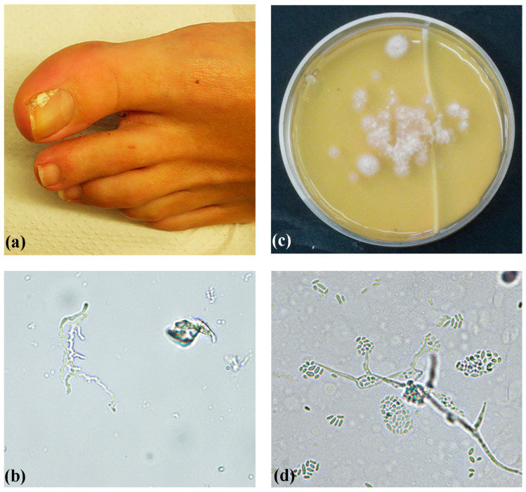
Figure 2.
A confirmed (repeated isolation and positive direct microscopy) toenail white superficial onychomycosis case in a 40-year-old woman, caused by a type 3 Acremonium egyptiacum isolate. (a) White area on the surface of the first toenail. (b) Direct microscopy of nail clippings showing a branching hyphal structure (original magnification 400×). (c) First isolation directly from nail clippings on Sabouraud dextrose agar, showing multiple A. egyptiacum colony forming units, after incubation at 32 °C for 8 days. (d) Microscopy showing slimy heads on phialides (original magnification 400×).

