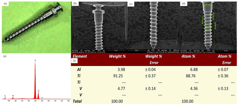
Figure 2.
Surface analysis of an unused OrthAnchor System implant. (a) Overview aspect seen by optical microscopy, 10×. (b) SEM aspect of the cervical half, Z1-Z3 areas. (c) SEM aspect of the apical half, Z2-Z4 areas. (d) SEM aspect of the implant, Z2-Z4 areas with measurements. (e) EDX analysis of the implant surface. (f) Percentage elemental analysis from the implant surface.

