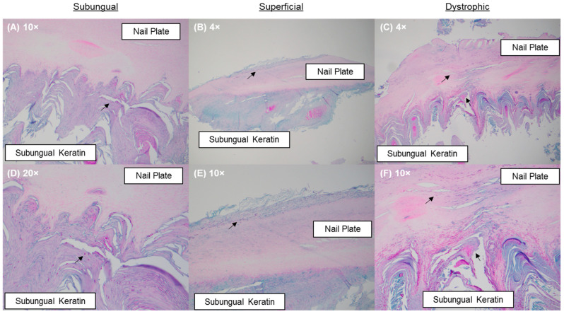
Figure 1.
PAS-stained nail specimens: (A,D) a subungual infection pattern evidenced by the presence of fungal elements in the subungual keratin; (B,E) a superficial infection pattern evidenced by the presence of fungal elements within the dorsal aspect of the nail plate; and (C,F) a dystrophic infection pattern evidenced by the presence of fungal elements in the nail plate and in the subungual keratin. Fungal elements are indicated by black arrows.

