Figure 2.
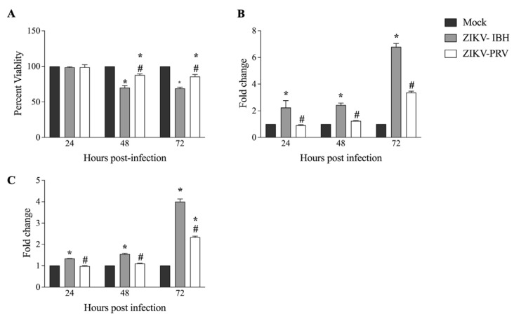
Significantly lower cytopathogenicity of Asian Lineage ZIKV compared with African lineage ZIKV. (A) ZIKV-PRV induces less cell death after infection in CHME-3 cells than ZIKV-IBH as measured by the MTS assay. Data presented as percent viability relative to mock infection (n = 6 biological replicates, Alpha 0.05). (B) ZIKV-IBH induces more necrosis after infection in CHME-3 cells than ZIKV-PRV as measured by quantification of extracellular LDH. Data presented as fold change to mock infection (n = 5 biological replicates, Alpha 0.05). (C) ZIKV-IBH displays more caspase-induced apoptosis in CHME-3 cells than ZIKV-PRV as measured by quantification of caspase 3/7 cleavage. Data presented as fold change to mock infection. (n = 12 biological replicates, Alpha 0.05). Significance from mock infection denoted by an asterisk (*) and significance between ZIKV lineages denoted by an octothorpe (#), each determined by t-test (p ≤ 0.05). All values are presented as mean ± SEM.

