Figure 3.

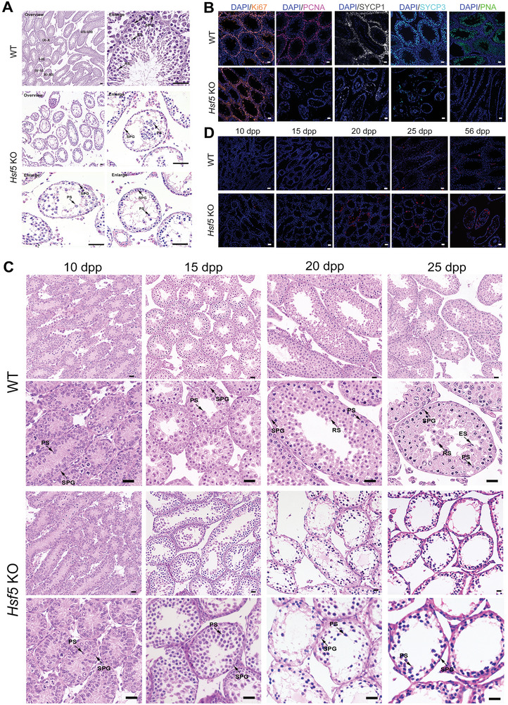
Hsf5 KO male mice exhibit infertility. A) Sections of testes from 8‐week‐old Hsf5 KO mice showing a decrease in spermatocytes and an absence of round and elongating/elongated spermatids compared with testes from WT mice. SPG, spermatogonium; PS, primary spermatocyte; RS, round spermatid; ES, elongating/elongated spermatid (N = 3 biologically independent WT mice and KO mice; scale bars, 125 µm). B) Immunofluorescence staining of spermatogenic cell markers in testes from WT and Hsf5 KO mice (N = 3 biologically independent WT mice and KO mice; blue, DAPI; orange, Ki67; pink, PCNA; grey, SYCP1; sky blue, SYCP3; green, PNA; scale bars, 125 µm). C) Histological analysis of testes from Hsf5 KO mice of different ages. H&E staining revealed the absence of RS and ES in Hsf5 KO testes at 20 dpp. SPG, spermatogonium; PS, primary spermatocyte; RS, round spermatid; ES, elongating/elongated spermatid (N = 3 biologically independent WT mice and KO mice; scale bars, 40 µm). D) TUNEL staining of apoptotic germ cells in WT and Hsf5 KO testis sections of different ages (N = 3 biologically independent WT mice and KO mice; blue, DAPI; red, TUNEL; scale bars, 125 µm).
