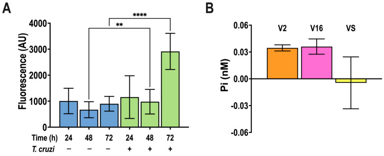
Figure 3.
α-SMA production by T. cruzi-infected NIH-3T3 cells, and polyP presence in trypomastigote extracellular vesicles. (A) NIH-3T3 cells were infected by T. cruzi cell-derived trypomastigotes (ratio 20:1) and α-SMA antibody (1:200) staining was quantified by fluorescence microscopy as a marker of myofibroblast differentiation at different times post-infection. Values are means ± S.D., n = 3, ** p < 0.01, **** p < 0.0001, ANOVA with Turkey’s multiple comparisons test. (B) PolyP extracted from large (V2) and small (V16) extracellular vesicles, and vesicle free supernatant (VS) of 1 × 10⁸/mL cell-derived trypomastigotes incubated for 18 h, at pH = 5.0, detected by the PPX method, and expressed in Pi units.

