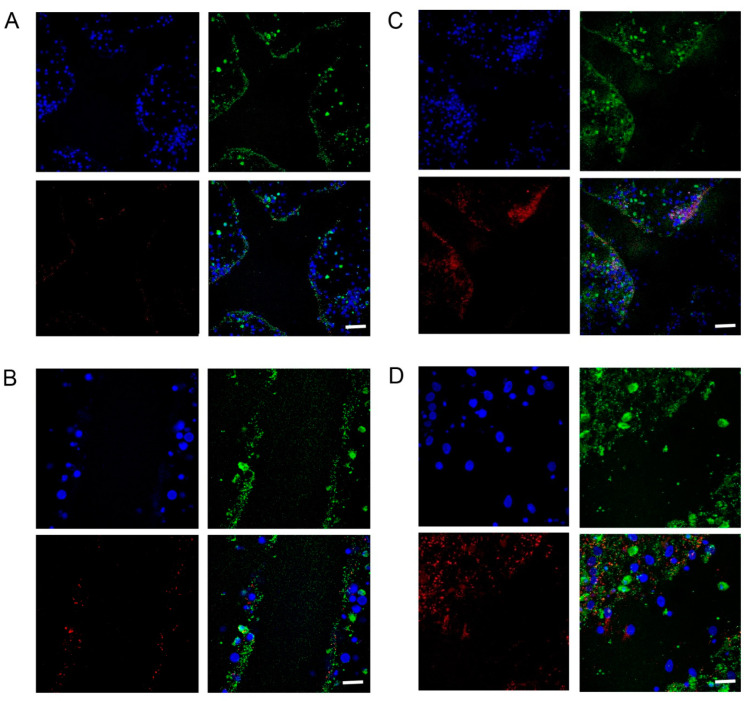
Figure 5.
Immunofluorescent staining shows that the density of cell nuclei (blue), synapsin-I (red) and beta-III-tubulin (green) increased with D/L-BHB treatment 24 h after injury. (A,B) In the control, fewer cell nuclei were visible around the regeneration site, and there was a lower density of synapsin-I and beta-III-tubulin. (C,D) With the BHB treatment, more cell nuclei were visible around the regeneration site, and there was a higher density of synapsin-I and beta-III-tubulin, compared to the control. Scale bar is 100 μm (A,C); 50 μm (B,D).

