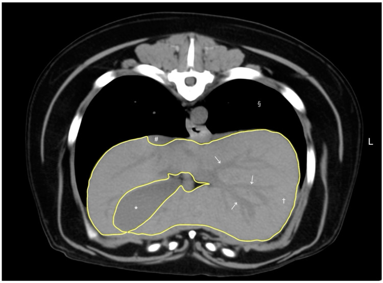
Figure 1.
A representative image of abdominal CT transverse pre-contrast images using CT liver volumetry in dogs. The window width and window level were adjusted to 350–400 HU and 40 HU, respectively. The liver segmentation was manually traced as a region of interest (ROI, yellow line). Hepatic vessels (white arrows) within the hepatic parenchyma (†) were included in the ROI, whereas the gallbladder (*), visible liver lobe fissures, and hepatic vessels outside the hepatic parenchymal margin were excluded. The caudal vena cava (#) and pulmonary parenchyma (§) were also noted.

