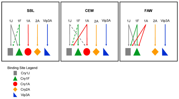
Figure 6.
Models depicting the binding site relationships between Cry1JP578V and the various proteins evaluated in each insect. Solid lines indicate complete binding site sharing and dashed lines indicate partial binding site sharing. Binding sites are represented by different geometric shapes and proteins are indicated the letters and numbers. In SBL BBMVs, Cry1JP578V only shares binding sites Cry1Fa but not with Cry2A.127 and Vip3A. In CEW BBMVs, Cry1JP578V, Cry1Fa and Cry1A.88 shared binding sites, while Cry2A.127 and Vip3A have completely independent binding sites. In FAW BBMVs, Cry1JP578V shares binding sites with Cry1A.88 and Cry1Fa, but also has an independent binding site that was not shared with Cry2A.127 and Vip3A.

