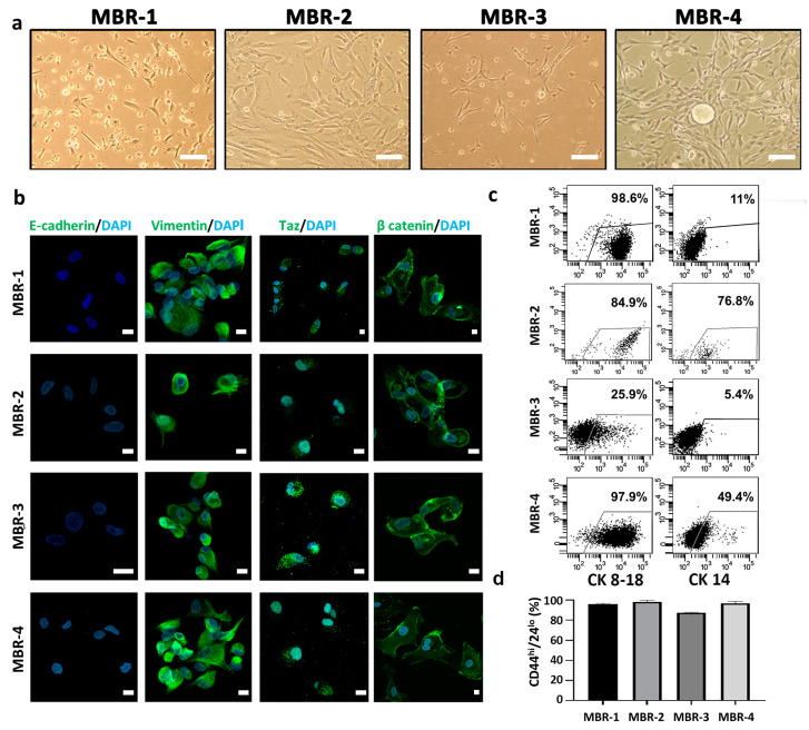
Figure 4.
Isolation and characterization of breast cancer patient-derived metastatic cells (PDMCs). (a) Representative phase-contrast images of PDMCs from ascitic fluid (MBR-1, MBR-2) or pleural effusion (MBR-3, MBR-4) cultured in serum-free conditions. Scale bar, 100 µm. (b) CLSM analyses of PFA-fixed PDMCs stained for E-cadherin, vimentin Taz and beta-catenin (green); DAPI was used to counterstain nuclei (light blue). Several fields were observed for each condition and representative images are shown. Scale bars, 10 µm. (c) Dot-plots showing luminal CK8-18 and myoepithelial CK14 expression in PDMC lines. (d) Bar chart reporting percentages of CD44highCD24−/low phenotype in MBR-1, MBR-2, MBR-3 and MBR-4 lines obtained from ascitic fluid or pleural effusion of breast cancer metastatic patients. Percentages, referring to CD44highCD24−/low positive cells, were determined by setting the gate on the isotype control from at least two independent FACS stainings.

