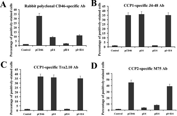
FIG. 6.
Detection of conformational changes in N-terminal CCP1 and CCP2 domains by antibody staining. CHO cells were transfected with plasmids expressing full-length or truncated forms of CD46 as described for Fig. 5A, and 24 hours later, cells were stained with indicated antibodies (Ab). Staining of primary antibodies was detected by Alexaflour-488-conjugated antibody and flow cytometry. All stainings were done in triplicate in two independent experiments. Error bars indicate standard deviations.

