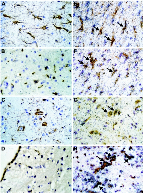
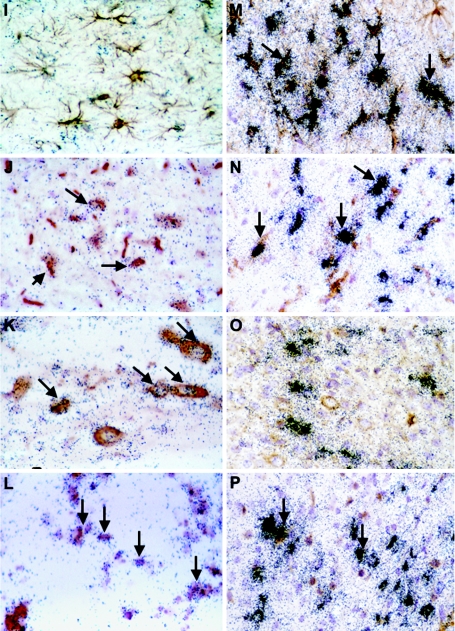
FIG. 3.
Cellular localization of IRF-7, IRF-9, and LCMV-NP mRNA in brain. WT mice were injected i.c. with saline or 250 PFU LCMV (ARM), and brains were removed at day 6 for in situ hybridization and immunohistochemistry. Eight-micron-thick paraffin-embedded sections were hybridized with 33P-labeled cRNA probes transcribed from linearized IRF-7, IRF-9, or LCMV-NP RPA plasmids and then processed for immunohistochemistry for cell-specific markers to identify CD3+ T cells, microglia, neurons, and astrocytes as outlined in Materials and Methods. Slides were coated with photographic emulsion, developed after 2 weeks, and visualized using bright-field microscopy. Photomicrographs of control (A to D) and day 6 LCMV-infected (E to P) brain sections hybridized for IRF-7 (E to H), IRF-9 (I to L), and LCMV-NP (M-P) mRNA and stained for astrocytes (A, E, I, M), microglia (B, F, J, N), neurons (C, G, K, O), and CD3+ T cells (D, H, L, P) are shown. IRF-7 hybridization was localized to astrocytes (E; arrows), microglia (F; arrows), neurons (G; arrows), and CD3+ T cells (H; arrows), while mainly microglia (I; arrows), neurons (K; arrows), and CD3+ T cells (L; arrows) expressed IRF-9 mRNA. Strong LCMV-NP hybridization was seen in astrocytes (M; arrows), microglia (N; arrows), some neurons (O), and CD3+ T cells (P; arrows).


