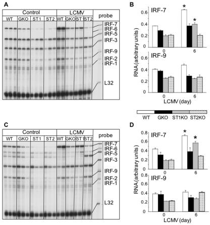
FIG. 6.
IRF mRNA levels in spleen (A, B) and liver (C, D) of uninfected (Control) and LCMV-infected WT, IFN-γ (GKO), STAT1 KO (ST1), and STAT2 KO (ST2) mice. Mice were injected i.c. with saline or 250 PFU LCMV (ARM), poly(A)+ RNA was isolated from the spleen and liver 6 days after injection, and 2 μg was analyzed by RPA as outlined in Materials and Methods. (A, C) Autoradiographs showing that IRF-2, -3, -5, -7, and -9 mRNA transcripts were constitutively present in normal, uninfected (Control) spleen and liver and additionally showing IRF-6 in livers of WT and GKO mice. In STAT1 KO and STAT2 KO animals, IRF-2, -3, -5, -7, and -9 mRNA transcripts were also constitutively expressed in spleen and liver, but the levels of IRF-7 and IRF-9 mRNAs were lower than those of the wild type. (B, D) Densitometric analysis of each lane was performed on scanned autoradiographs using NIH Image software (version 1.31). In both spleen (B) and liver (D), the level of IRF-7 mRNA increased significantly (*P < 05) after LCMV infection in wild-type and STAT1 KO mice compared with that of uninfected controls.

