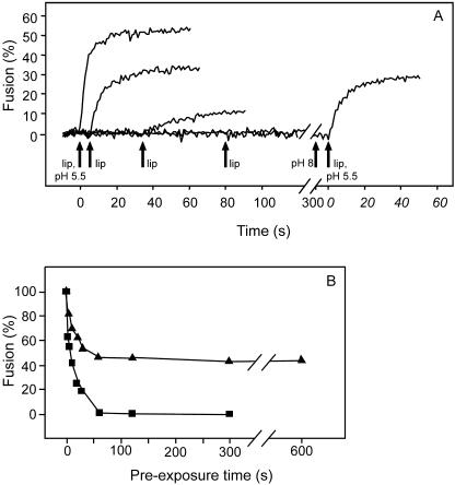
FIG. 1.
Effects of acidification, neutralization, and reacidification on SFV fusion with LUVs. Fusion of pyrene-labeled SFV with LUVs was measured on-line as the decrease of the pyrene-excimer fluorescence at 480 nm (33). The fusion scale was set such that the initial excimer fluorescence at 480 nm represented 0% fusion and the residual fluorescence, after addition of the detergent octaethyleneglycol-monododecyl ether to a final concentration of 10 mM (representing infinite dilution of the fluorophore), corresponded to 100% fusion. Panel A presents a compilation of multiple measurements in which virus (1 μM), in a continuously stirred final volume of 0.7 ml HNE buffer (pH 7.4) in a thermostated quartz cuvette (37°C), was acidified to pH 5.5 by the injection of a small, pretitrated volume of 0.1 M MES (morpholinoethanesulfonic acid) and 0.2 M acetic acid at 0 s. Subsequently, PC-PE-Chol-SPM (1:1:1.5:1) LUVs (100 μM) were added at 5 s, 35 s, and 80 s (arrows). For a control, a mixture of virus and LUVs was acidified to pH 5.5 at 0 s (first curve on the left). At 300 s, the pH of the virus suspension was neutralized to pH 8 by the injection of a small volume of 0.1 M NaOH (second arrow from the right), after which LUVs were added, and the mixture was acidified again to pH 5.5 with a pretitrated volume of 0.1 M MES and 0.2 M acetic acid (outer arrow on the right). lip, liposomes (LUVs). For panel B, virus was acidified as described for panel A, and at the indicated time points, PC-PE-Chol-SPM LUVs were added (squares) or the mixture was neutralized first, after which LUVs were added and the mixture was reacidified to determined the recovery of fusion activity of the virus (triangles).

