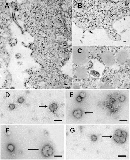
FIG. 3.
Electron micrographs of BHK cells infected with the mutant KK391/392. As seen in panel A, this mutant produces wild-type levels of nucleocapsids seen within the cytoplasm and found associated with cell membranes. This mutant, however, produces long tubes of membrane containing multiple associated cores. Seen in panel B are virus particles containing 2, 3, and 6 cores budding from the cell surface. In panel C, a large bolus of virus cores is seen budding within an internal vesicle, surrounded by membranes associated with virus cores. In panels D to G, negatively stained purified mutant virus is seen forming conjoined particles containing 2 (D), 3 (E, F), and 4 lobes (G). Bars, 100 nm.

