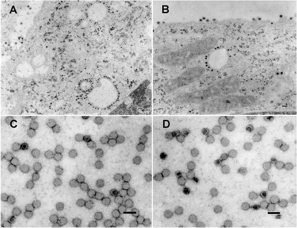
FIG. 4.
Electron micrographs of BHK-grown F391 mutant. Panels A and B are representative of infected BHK cells. In panel B, many particles are seen budding directly from the cell membrane. In panels C and D, negatively stained gradient-purified virus from BHK cells display normal spherical virus particles with a few empty particles visible. Bars, 100 nm.

