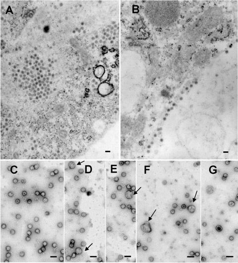
FIG. 5.
Electron micrographs of FF391/392 mutant-infected insect U4.4 cells. In panel A, large internal vesicles containing matured virus are visible, while in panel B, particles are seen budding from the plasma membrane. Both panels A and B show particles with a wild-type appearance. Panels C to G display negative stains of gradient-purified mutant virus. Arrows in panels D to F highlight a few larger particles that are formed by this mutant in the insect cell host. Compared with mutant virus particles produced from BHK cells (Fig. 2D and E), the virus morphology of the FF391/392 mutant grown in U4.4 cells is more similar to the wild-type virus (Fig. 1D). Bars, 100 nm.

