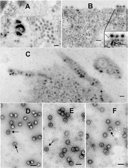
FIG. 6.
Electron micrographs of insect U4.4 cells infected with the mutant KK391/392. In panel A, a cytoplasmic vesicle containing matured virus is shown. In panel B, the inset shows budding virus particles with visible membrane stalks (arrow), indicative of an aberrant membrane fission process. Panel C demonstrates long tubes of multicored membrane appendages also seen produced by this virus grown in BHK cells (Fig. 3A, B, and C). Negative stains of membrane-purified virus reveal a different phenotype than that seen for this mutant grown in BHK cells (Fig. 3D to G). (D to F) Virus grown in U4.4 cells appears pocked and fragile (arrows). Bars, 100 nm.

