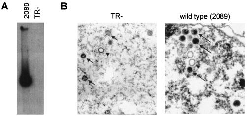
FIG. 2.
(A) Gardella gel analysis of viral supernatants. One milliliter of supernatant from induced 293/TR− or 293/2089 cells was ultracentrifuged, and the pellet was loaded onto a Gardella gel. No viral DNA could be detected in the TR− virus pellet. The 2089 virus pellet gave rise to a strong signal, as expected. (B) Electron micrographs of induced 293/TR− and 293/2089 cells. Cells were harvested at day 3 postinduction and fixed, and thin sections were prepared. Numerous A capsids (arrowheads) and B capsids (black short arrows) are visible within the nuclei of induced 293/TR− cells. In contrast, induced 293/2089 cells contain mainly mature virions with packaged EBV genomes (black long arrows).

