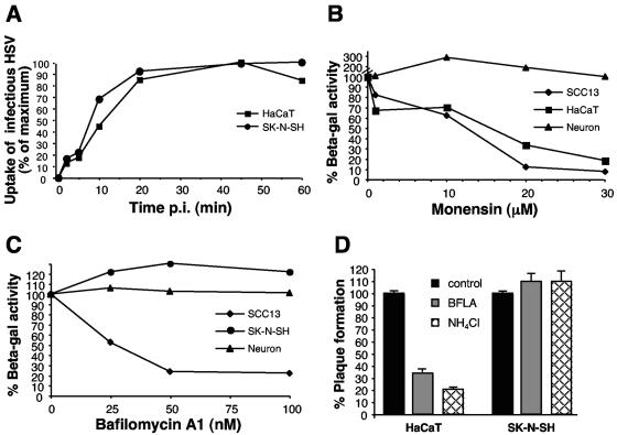
FIG. 1.
Effect of lysosomotropic agents on HSV infection of neurons and keratinocytes. (A) Uptake of infectious HSV from the cell surface. HSV-1 KOS K26GFP was bound to HaCaT keratinocytes or SK-N-SH neuroblastoma cells for 1 h at 4°C (MOI of 0.5). Cells were washed with PBS and incubated at 37°C, and extracellular virus was inactivated by acid treatment at the indicated times. At 8 hpi, cells were fixed and random fields of ∼1,000 cells in total were evaluated per sample. Cell number was determined by nuclear staining with DAPI, and infected, GFP-positive cells were counted. Maximum infectivity was set to 100%. (B, C) Effect of lysosomotropic agents on HSV-induced gene expression. Cells were pretreated with the indicated concentrations of agent for 1 h. Cells were infected with the lacZ+ HSV-1 strain KOS 7134 at an MOI of 1 for 7 h in the continued presence of agent. (D) Effect of lysosomotropic agents on HSV plaque formation. Cells were pretreated with 100 nM bafilomycin or 50 mM ammonium chloride for 1 h. HSV-1 KOS was added for 6 h at 37°C in the presence of the agent. The monolayers were washed and then incubated in normal medium for an additional 16 h. Plaques were visualized by immunoperoxidase staining with anti-HSV polyclonal sera. Each point represents the mean of quadruplicate wells.

