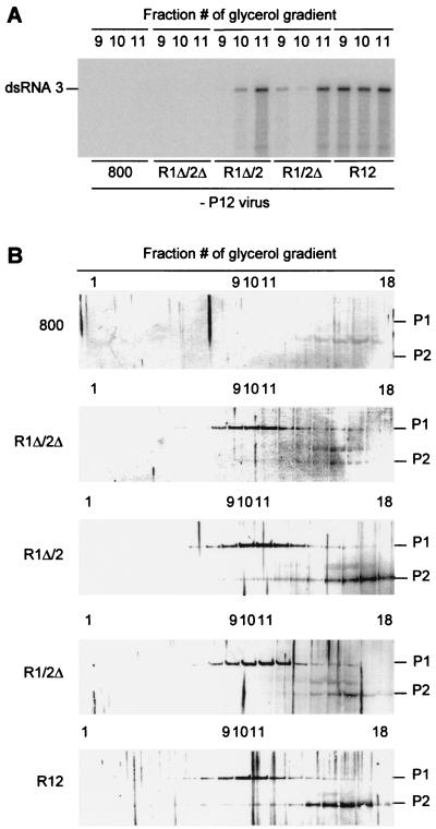
FIG. 5.
Accumulation of replicase proteins and RdRp activity in agroinfiltrated leaves. Leaves were infiltrated with A. tumefaciens suspensions containing the empty T-DNA vector or constructs R1Δ/2Δ, R1Δ/2, R1/2Δ, or R12 as indicated at the bottom of panel A and in the left margin of panel B. Two days after infiltration, the leaves were homogenized, after which RdRp was solubilized from the 30,000 × g membrane fraction and sedimented in a glycerol gradient. (A) In vitro RdRp assay. Samples from fractions 9, 10, and 11 of the glycerol gradients were used in RdRp assays with plus-strand AMV RNA 3 as a template (fraction 1 is at the bottom of the gradient). Radiolabeled products were analyzed by gel electrophoresis. The position of double-stranded RNA 3 is indicated in the left margin. (B) Western blot analysis of the protein composition of glycerol gradient fractions. Fractions 1 (bottom) to 18 (top) were analyzed with antisera to P1 and P2 proteins. The positions of P1 and P2 are indicated in the right margin.

