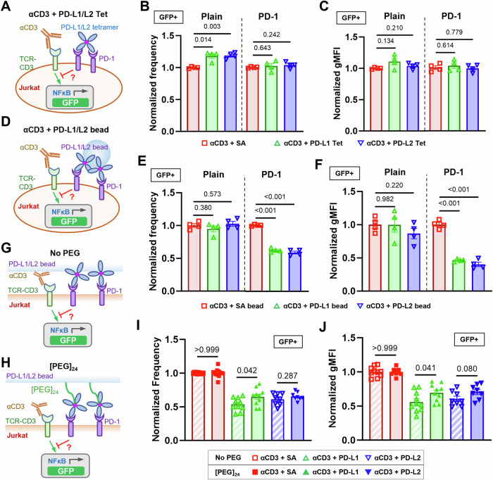
Fig. 1. The inhibitory function of PD-1 on Jurkat cells is enhanced by mechanical support of PD-Ligands.
A Schematics of stimulating NFκB::eGFP reporter Jurkat cells with soluble anti-CD3 and soluble PD-ligand tetramers. B, C Quantification of GFP expression for condition in (A). n = 4 for all conditions pooled from two independent experiments. D Schematics of stimulating NFκB::eGFP reporter Jurkat cells with soluble anti-CD3 and PD-ligand-coated beads. E, F Quantification of GFP expression for condition in (B). n = 4 for all conditions pooled from two independent experiments. Schematics of stimulating NFκB::eGFP reporter Jurkat cells with soluble anti-CD3 and PD-Ligands coated beads without (G) or with (H) [PEG]24 spacer arm. I, J Quantification of GFP expression for conditions in (G, H). n = 10, 10, and 8 for SA, PD-L1, and PD-L2, respectively, pooled from 5 independent experiments. Normalized frequency (B, E, I) and normalized geometric mean fluorescence intensity (gMFI) (C, F, J) were calculated as (sample—averaged background)/(anti-CD3 control—averaged background) and presented as mean ± SEM. Numbers on graphs represent p values calculated from two-tailed student t test. Source data are provided in Source Data file.

