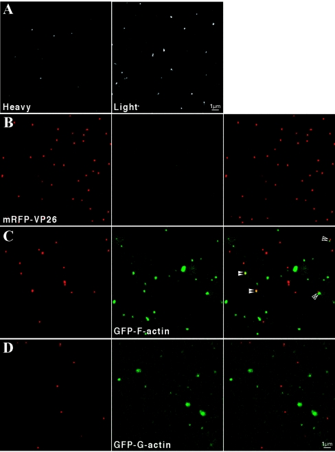
FIG. 4.
Detection of filamentous and nonfilamentous GFP-actin in single virions. Becker virions produced from the infection of heterogeneous GFP-F-actin expressing cells (line 2) were purified on a linear tartrate gradient (5 to 20%). (A) Particles isolated from the heavy (left) or light (right) bands were combined with an equal volume of glycerol and imaged by confocal microscopy. Particles from the light band exhibited increased fluorescence relative to those from the heavy band. (B) Recombinant PRV 180 virions, containing an mRFP-VP26 (red-capsid) fusion and isolated from infected PK15 cells, exhibited red fluorescence (left) but no detectable green fluorescence (middle). (C) A similar infection and analysis of homogenous GFP-F-actin-expressing cells (line 74) revealed the green fluorescence overwhelmingly localized to light particles (right panel, merged red and green). Occasionally, GFP-F-actin was observed to colocalize with red capsid puncta (filled arrowheads) or to be juxtaposed to red capsid (open arrowheads) in heavy particles. (D) PRV 180 infection of GFP-G-actin cells (line 52) also revealed that green fluorescence predominantly localized to light particles.

