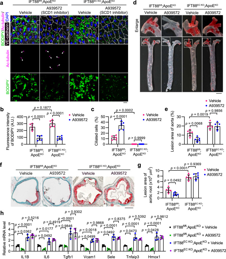
Fig. 6. SCD1 inhibition attenuates atherosclerosis progression in an endothelial cilia-dependent manner.
a–c En face immunofluorescence images (a) and quantifications of BODIPY staining (b) and ciliation (c) of aortic arch VECs from IFT88EC KO;ApoEKO and littermate ApoEKO mice intravenously injected with the SCD1 inhibitor A939572 (5 mg/kg body weight/2 days) or vehicle for 4 weeks after an 8-week HFD feeding (n = 6 mice). Scale bar, 10 μm. d Representative images of Oil Red O (ORO) staining of atherosclerosis lesions on the aorta of IFT88EC KO;ApoEKO and littermate ApoEKO mice treated as described in (a). Boxed areas are enlarged in the top panel. Scale bar (for enlarged images), 1 mm. e Quantification of atherosclerotic lesions shown in (d) (n = 6 mice). f Representative ORO staining in the aortic root of IFT88EC KO;ApoEKO and littermate ApoEKO mice treated as described in (a). Scale bar, 500 μm. g Quantification of atherosclerotic lesions shown in (f) (n = 6 mice). h Transcriptional levels of IL1B, IL6, Tgfb1, Vcam1, Sele, Tnfaip3, and Hmox1, in mice treated as described in (a) (n = 4 mice). Data are presented as mean ± SEM. Statistical significance was determined by two-way ANOVA with post hoc analysis. Source data are provided as a Source Data file.

