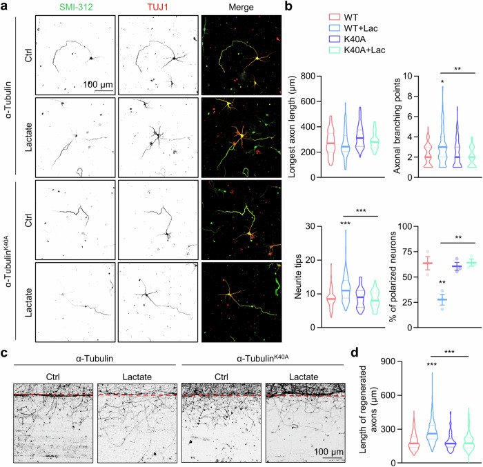
Fig. 6. α-Tubulin lactylation promotes axon outgrowth and branching.
a Representative images of hippocampal neurons at DIV3. The hippocampal neurons were transfected with α-tubulin or α-tubulin K40A mutant. The neurons were treated with 30 mM lactate for 72 h, and stained with anti-SMI-312 (green) and anti-Tuj-1 (red) antibodies. b Quantitative analysis of the longest axon length, number of axonal branching points, number of neurite tips, and percentage of polarized neurons in cultured neurons in (a). For WT, WT + Lac, K40A, and K40A + Lac, n = 66, 74, 54, and 62 neurons, respectively, from 3 experiments. One-way ANOVA, for axonal branching points, WT + Lac vs WT, p = 0.0265; K40A + Lac vs WT + Lac, p = 0.0024. For neurite tips, WT + Lac vs WT, p < 0.0001; K40A + Lac vs WT + Lac, p < 0.0001. For polarized neurons, WT + Lac vs WT, p = 0.0039; K40A + Lac vs WT + Lac, p = 0.0035. c Representative images of axon regeneration. The cortical neurons were infected with lentiviruses encoding α-tubulin or α-tubulin K40A mutant. The axons were severed after being treated with or without 30 mM lactate for 4 h at DIV7 and axon regeneration was assessed 1 day later. d Quantitative analysis of data in (c). For WT, WT + Lac, K40A, and K40A + Lac, n = 222, 221, 175, and 159 axons, respectively, from 3 experiments. One-way ANOVA, WT + Lac vs WT, p < 0.0001; K40A + Lac vs WT + Lac, p < 0.0001. Data are shown as mean ± SEM. *p < 0.05, **p < 0.01, ***p < 0.001. Source data are provided as a Source Data file.

