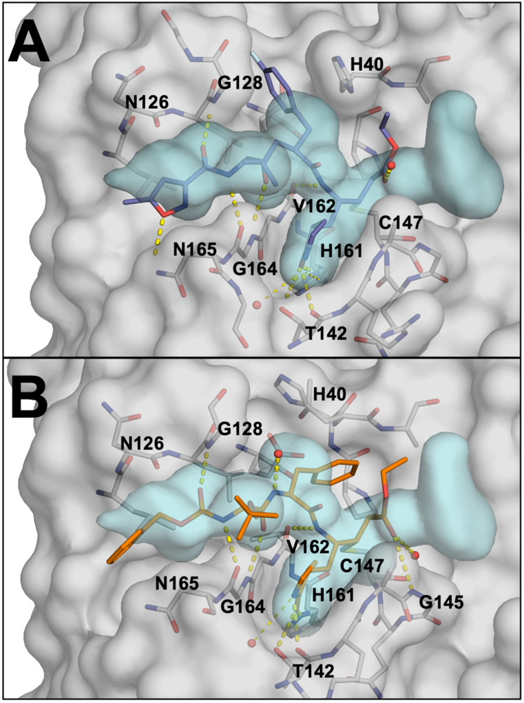
Figure 6.
(A) Rupintrivir (AG7088), in slate blue, covalently bound within the active site of EV68-3C protease in grey (PDB: 7L8H). Hydrogen bonds between rupintrivir and the protein are highlighted as yellow dashed lines. The substrate envelope is shown in light blue (B) SG-85, in orange, covalently bound within the active site of EV68-3C protease (PDB: 3ZVF).

